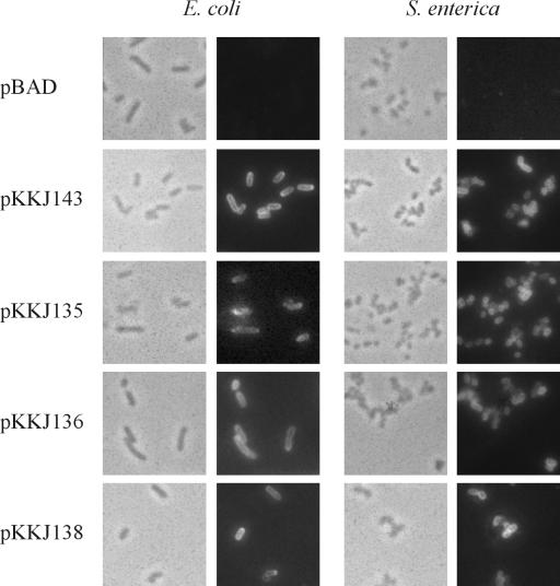
FIG. 4.
Immunofluorescence microscopy showing surface presentation of Ag43 on recombinant E. coli HEHA16 cells and S. enterica SL5325 cells. Phase contrast microscopy (the left panels on each side) and fluorescence microscopy (right panels on each side) were performed. To detect the presence of Ag43 on the cell surface, a polyclonal rabbit serum raised against the α-subunit of Ag43 was used, and this was detected by FITC-labeled anti-rabbit immunoglobulin serum.

