
Figure 2.
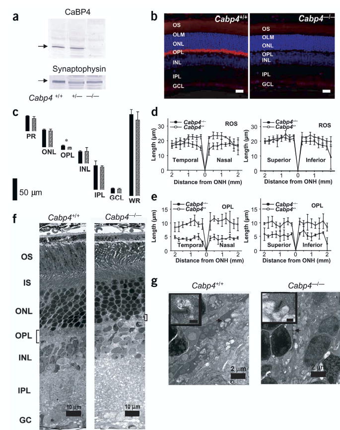
Characterization of CaBP4 knockout mice. (a) Immunoblotting of retinal extracts from Cabp4+/+, Cabp4+/− and Cabp4−/− mice probed with anti-CaBP4 or anti-synaptophysin (control). (b) Immunolocalization of CaBP4 in the retina of Cabp4+/+ mouse (left). Lack of CaBP4 immunoreactivity in the retina of Cabp4−/− mouse (right). Scale bars, 20 μm. Nuclei are visualized by staining with Hoechst 33342 dye (blue). (c) The thickness of individual retinal layers from 8-to 10-week-old Cabp4+/+ (black bars) and Cabp4−/− mice (gray bars) measured at 1.25 mm inferior from the optic nerve head. *P < 0.01. GCL, ganglion cell layer; INL, inner nuclear layer; IPL, inner plexiform layer; ONL, outer nuclear layer; OPL, outer plexiform layer; PR, photoreceptor outer and inner segments; WR, whole retina. (d,e) Rod outer segment (ROS; d) and outer plexiform layer (ORPL; e) thickness (in micrometers) plotted as a function of the retinal location (in millimeters) from the optic nerve head. (f) Montage of cross-sections through the retinas of 2-month-old mice analyzed by transmission electron microscopy. The outer plexiform layer is thinner in Cabp4−/− mice than it is in Cabp4+/+ mice. IS, inner segment; OS, outer segment. (g) Higher magnification of a cross-section through the outer plexiform layer. Photoreceptor terminals are present in the outer plexiform layer of Cabp4+/+ mice (arrow), but fewer and altered terminals are observed in Cabp4−/− mice. A synaptic ribbon is shown at higher magnification (inset; scale bars, 0.2 μm).