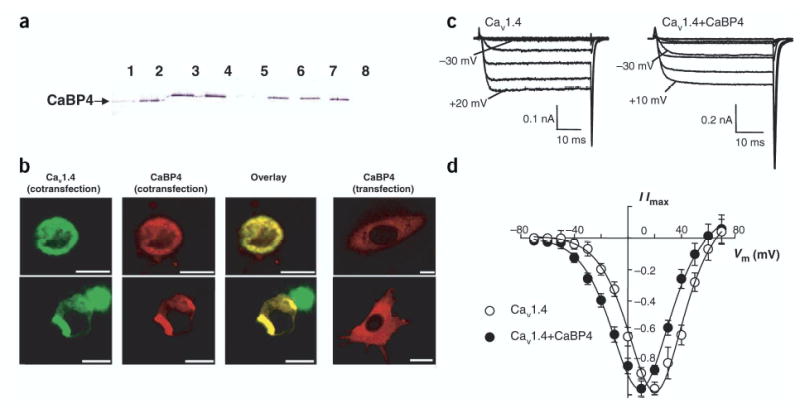
Figure 7.

CaBP4 interacts with and modulates Cav1.4. (a) Affinity chromatography of purified recombinant mCaBP4 on Ca1.4vα1 column. The eluted fractions were probed with anti-CaBP4. Lanes 1–4, elution with 3 mM EGTA; lane 5, final elution with 3 mM EGTA; lanes 6–8: further elution with 0.1 M glycine buffer, pH 2.1. (b) Colocalization of CaBP4-DsRed2 with Cav1.4-GFP in HEK293 cells. Confocal images of HEK293 cells transfected with mCaBP4-DsRed2 and Cav1.4–GFP (left three panels) or transfected with mCaBP4-DsRed2 alone (right panel). The yellow color indicates colocalization of the overexpressed fusion proteins. Scale bars, 12.5 μ) m. (c) Modulation of Cav1.4 activation by CaBP4 in transfected HEK293T cells. Whole-cell Ca2+ currents recorded in cells transfected with Cav1.4 subunits (α1 1.4, β2A, α2δ) alone (left) or cotransfected with CaBP4 (right). Shown are representative traces of Ca2+ currents evoked by 50-ms steps from a holding voltage of −80 mV to various test voltages. Extracellular recording solution contained 20 mM Ca2+ and intracellular solution contained 5 mM EGTA. CaBP4 enhanced activation of Ca2+ currents at negative voltages as indicated. (d) Current-voltage relationship from cells transfected with Cav1.4 alone (○) or cotransfected with CaBP4 (•). For each test voltage (Vm), I/Imax represents the current amplitude measured at 45 ms normalized to the maximal current amplitude obtained in the series (mean ± s.e.m.; n = 7–10).
