
Fig.4.
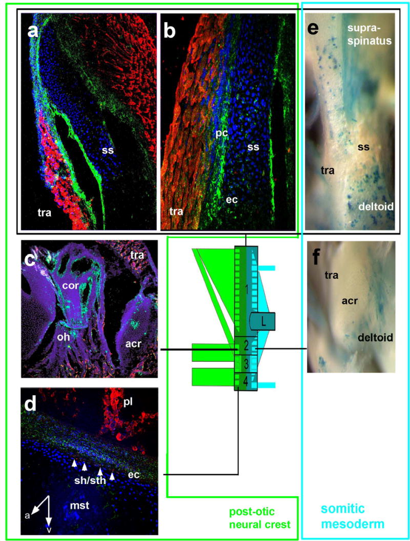
Dual neural crest and mesodermal origins of the endochondral shoulder girdle. Green box PONC GFP+ green (a,b) components, turquoise box mesodermal LacZ+ blue (e) components of the scapular spine (box 1, a,b,e), the acromio-coracoid (box 2, c,f,) and sternum (box4,d). Inside the scapular spine (ss, a,b) GFP+ neural crest cells are found to form endochondral skeleton (ec) and perichondrium (pc) exactly at the places where the trapezius (tra) carrying crest connective tissue is attached. Conversely, blue mesodermal muscle fibres are attached to punctuate areas of the posterior scapular spine (e) and acromion (f). Note that areas of trapezius attachment in mice with mesodermal labeling are white, i.e. unlabelled, showing that neural crest and mesodermal muscle attachment systems do not mix with each other. coracobranchial muscle fibres (Sh/sth in d) are inserted by the endochondral PONC component (white arrows) onto the mesodermal sternum (mst). A anterior, v ventral, pl pleural muscles.