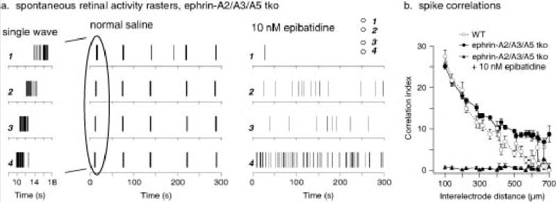
Fig. 5.

Spontaneous retinal waves in ephrin-A2/A3/A5 mutant retinas. (a) Spike rasters of spontaneous retinal activity from an ephrin A2/A3/A5 triple knockout (tko) mouse at P4. Activity at single ganglion cells occurs in bursts followed by long periods of silence. Recording on a multielectrode array shows that these bursts occur across the retina in waves. Relative electrode positions (1 – 4) for the example neuron recordings are illustrated in the inset of the right panel. Closer examination of a typical wave (left panel, 10 sec) shows it traveling from electrode 4 to 1 at 140 μm/sec. In contrast, the activity of the same neurons in the presence of 10 nM epibatidine shows no waves. (b) Correlation indices were calculated for all pairs of neurons recorded from a P4 wild-type retina (white circles), a P4 ephrinA2/A3/A5 tko retina (black circles), and an epibatidine treated ephrin-A2/A3/A5 tko retina (black triangles). These are plotted against the distance between the electrodes from which the neuron’s spikes were recorded. In both the wild-type and ephrin A2/A3/A5 tko retinas, wave activity led to high correlation indices at small distances and lower ones at larger distances. Ephrin A2/A3/A5 tko retinas had a higher level of correlation at the largest distances in the array (approximately 800 μm) than the wild-type retinas, due to waves that were somewhat broader. Epibatidine, which eliminated the wave activity but did not eliminate spike activity in the ephrin A2/A3/A5 tko retina, abolished correlated activity at all distances. Wild-type n=3 retinas, ephrin-A2/A3/A5 n=3 retinas.
