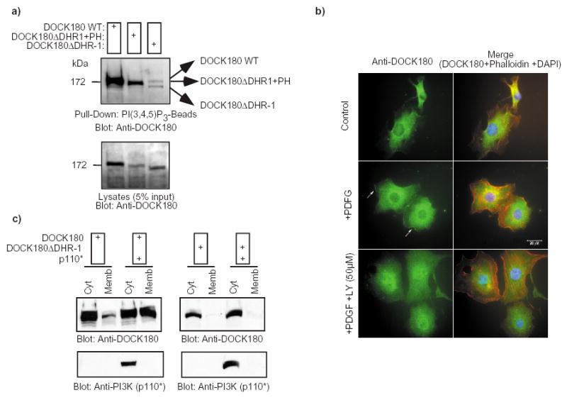
Fig. 5. The DHR-1 domain displays lipid binding activity in vivo.

a) The DOCK180 DHR-1 mutant fails to interact with PtdIns(3,4,5)P3. DOCK180 WT, DOCK180 DHR-1 or DOCK180 DHR-1+PH (see Fig. 7 and supplemental information, Fig. S1) were expressed in LR73 cells. Cell lysates were subjected to a pull-down by PtdIns(3,4,5)P3-beads. Bound proteins were detected by immunoblotting with an anti-DOCK180 antibody (C19). The lower panels demonstrate the expression levels of the exogenous DOCK180 proteins. b) Endogenous DOCK180 localizes to the plasma membrane in response to PDGF stimulation in a PtdIns 3-kinase dependent manner. Serum-starved NIH3T3 cells were treated with either DMSO (top and middle panels) or 50 μM LY294002 for 30 min (bottom panels). Cells were subsequently left untreated (top panels) or stimulated with 10 ng/ml PDGF (middle and bottom panels) for 10 min prior to fixing. Cells in the left panels were stained with an anti-DOCK180 rabbit polyclonal antibody, while the panels on the right represent an overlay of the anti-DOCK180, rhodamine-phalloidin and DAPI stains. Cells were photographed at a 60X magnification. c) DOCK180 translocates to the membrane in response to PtdIns(3,4,5)P3 production in a DHR-1-dependent manner. HEK293T cells were transfected with the indicated plasmids. After 24 h, the cytosolic and membrane fractions were biochemically purified and the distribution of DOCK180, DOCK180 DHR-1 and p110* was analyzed by immunoblotting with the indicated antibodies.
