
Fig. 7. The PH domain of BMX/Etk can functionally replace the DHR-1 domain in DOCK180.
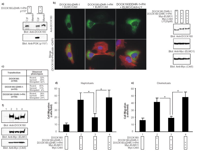
a) A chimeric DOCK180 protein (DOCK180 DHR-1+PH) in which the DHR-1 domain had been replaced with the PH domain of Bmx/Etk translocates to the membrane in response to PtdIns(3,4,5)P3 production. HEK293T cells were transfected with the indicated plasmids. After 24 h, the cytosolic and membrane fractions were biochemically purified and the distribution of DOCK180 DHR-1+PH and p110* was analyzed by immunoblotting with the indicated antibodies. b) The introduction of the PH domain of Bmx/Etk in the DHR-1 mutant rescues the cell polarization defects. Serum-starved LR73 cells expressing the indicated plasmids were detached and allowed to spread on fibronectin for 2 h. Cells were then analyzed as described in Fig 1a. When treated with LY294002, the cells were preincubated with the inhibitor for 30 min prior to plating and the inhibitor was left on the cells throughout the experiment. In the right panel, expression levels of the proteins were analyzed by immunoblotting the cell lysates with anti-DOCK180 (C19) and anti-Myc antibodies. c) Quantification of the effect on cell elongation by the DOCK180 DHR-1+PH chimeric protein. Cells were processed exactly as in (b) and several independent fields were photographed. The cells were visually inspected and scored for three phenotypes: 1- Round (attached and minimally spread cells), 2- Spread (clearly spread and flat cells) and 3- Elongated (elongated cells with a polarity). This is a representative experiment of three independent assays. d–e) The introduction of the PH domain of Bmx/Etk in the DHR-1 mutant rescues the cell migration defects. Serum-starved LR73 expressing the indicated plasmids were detached and subjected to haptotactic and chemotactic migration assays, as described in Fig. 1. Data represent mean +/- SD of a representative experiment performed in duplicate. *, P<0.001; one-way ANOVA. f) Expression levels of the transfected proteins were analyzed by immunoblotting of cell lysates with anti-DOCK180 and anti-Myc antibodies, as indicated.