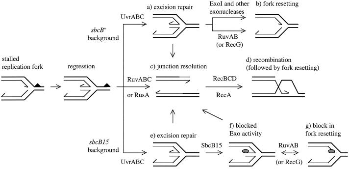
FIG. 1.
Effect of the SbcB15 protein on the repair of replication forks stalled at pyrimidine dimers. The upper part of the figure represents molecular events occurring in UV-irradiated wild-type (sbcB+) cells. The lower part pictures reactions occurring in the sbcB15 background. Reactions shown in the middle part of the figure are common to both backgrounds. The solid triangle and the shaded oval represent a pyrimidine dimer and the SbcB15 protein, respectively. The directions of DNA synthesis within replication forks are indicated by arrows. Details of the model are discussed in the text.

