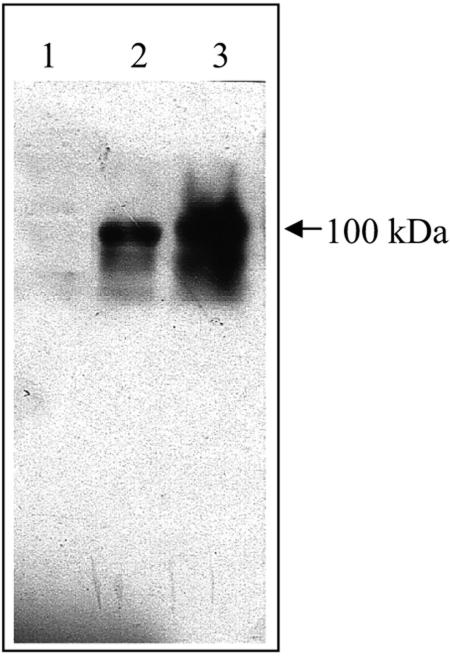
FIG. 3.
Western blot analysis of GP1 S-layer protein. Lane 1, control strain, B. thuringiensis 407 transformed with pHT315 plasmid; lane 2, B. thuringiensis 407 strain transformed with pHT-GP1 plasmid; lane 3, GP1 strain. Samples were subjected to SDS-PAGE, and the GP1 SL protein was detected by Western blotting with an anti-GP1 SL polyclonal antibody.

