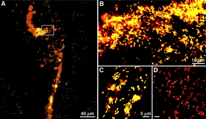
FIG. 3.
False color images of bacterial cells detected by FISH in egg capsules of E. fetida using both Cy3-LSB 145 and FL-Eub 338. (A) laser scanning confocal microscope image of a large aggregate representative of aggregates detected in the egg capsule albumin. (B to D) Yellow cells, Acidovorax; red cells, Eub 338 only. (B) Enlargement of the area in the box in panel A. (C) Acidovorax cells, showing morphology similar to the morphology of cells observed in ampullas of E. fetida nephridia. (D) Abundant cells labeled with only Eub 338.

