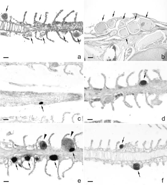
FIG. 2.
Detection of Chlamydiales bacteria in epitheliocystis of gills of barramundi (Lates calcarifer) and silver perch (Bidyanus bidyanus) by ICC and in situ RNA hybridization. (a) HE staining of gills of barramundi showing epitheliocystis cysts (arrows). (b) HE staining of lymphocystis cysts (arrows) in the skin of barramundi, clearly different in size and morphology from the cysts in the gills. (c and d) Some cysts in gills of barramundi stained clearly with antibodies raised against a membrane protein of Chlamydophila pneumoniae (c; arrow) and with antibodies raised against the lipopolysaccharide of Chlamydia trachomatis (d; arrow). (e and f) Abundant staining of cysts in gills of barramundi (e; arrows and arrowheads) and silver perch (f; arrows) by RNA ISH with a Chlamydiales-specific 16S rRNA oligonucleotide probe. Larger cysts in the gills of barramundi (e; arrowheads) stained less intensely than the smaller cysts (e; arrows). Bars: a and c to f, 10 μm; b, 100 μm.

