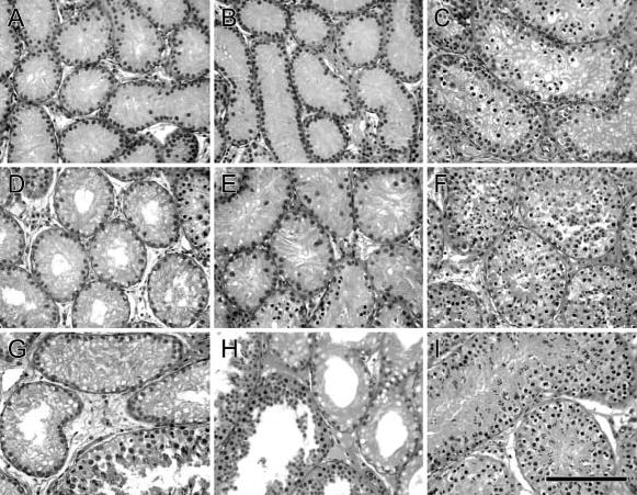
Figure 2.

Histological appearance of goat testis tissue at 2, 3, and 6 months after fractionated irradiation compared with testes from age-matched control animals. (A, D, G) Testis tissue from goats irradiated at 1 week of age analyzed at 2, 3, and 6 months after irradiation. (B, E, H) Testis tissue from goats irradiated at 5 weeks of age analyzed at 2, 3, and 6 months after irradiation. (C, F, I) Testis tissue from 3-, 4-, and 7-month-old control goats. Bar = 70 μm.
