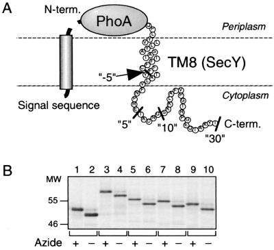
FIG. 1.
Signal sequence cleavage indicates the periplasmic localization of the N termini of the PhoA-TM8 model proteins. (A) Schematic representation of PhoA-TM8-Cn fusion proteins. The C-terminal ends for the constructs used (n = −5, 5, 10, and 30) are indicated by the black bars. (B) Demonstration of signal sequence-processed and unprocessed species. Strain AR5087 was transformed with either pCH312 (carrying PhoA; lanes 1 and 2), pCH309 (carrying PhoA-TM8-C30; lanes 3 and 4), pCH308 (carrying PhoA-TM8-C10; lanes 5 and 6), pCH307 (carrying PhoA-TM8-C5; lanes 7 and 8), or pCH306 (carrying PhoA-TM8-C−5; lanes 9 and 10). Cells were grown in M9 medium at 37°C, induced for the lac transcription for 10 min, and treated with (lanes with odd numbers) or without (lanes with even numbers) 0.02% NaN3 for 1 min. They were then pulse-labeled with [35S]methionine for 1 min, which was followed by a chase with unlabeled methionine for 1 min. Labeled proteins were immunoprecipitated with anti-PhoA serum, separated by SDS-PAGE, and visualized.

