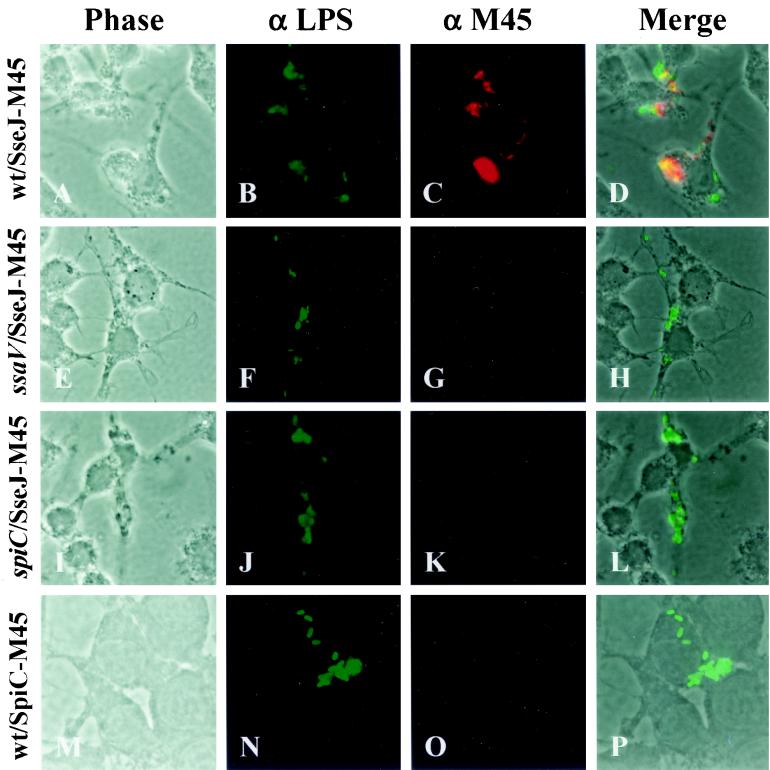
FIG. 3.
Translocation of epitope-tagged SPI2 effector proteins. RAW264.7 cells were infected with wild-type Salmonella serovar Typhimurium strain NCTC 12023 expressing SseJ-M45 (A to D), ssaV mutant strain HH109 expressing SseJ-M45 (E to H), spiC mutant strain EG10128 expressing SseJ-M45 (I to L), and wild-type strain NCTC 12023 expressing SpiC-M45 (M to P). Sixteen hours postinfection, cells were fixed, permeabilized, and stained for bacteria (α LPS, green) and epitopes (α M45, red). Stained cells were then examined by fluorescence microscopy and photomicrographs were taken of phase contrast, green fluorescence, and red fluorescence. Merged images, in which yellow represents colocalization of green and red fluorescence, were generated with Metavue imaging software (Universal Imaging Corporation).

