

Abstract
1. Intracellular records were obtained from giant reticulospinal cells (Müller cells) in the brain of adult lamprey. The cells had maximum resting potentials of -80 mV and action potentials with overshoots of 30 mV. Input resistances varied from 2 to 8 MΩ.
2. Individual spontaneous excitatory and inhibitory synaptic potentials (e.p.s.p.s and i.p.s.p.s) were observed, as well as occasional high frequency bursts of excitatory potentials. Much of the spontaneous synaptic activity could be eliminated by elevating the Ca2+ concentration in the bathing solution to 10-15 mM, suggesting that the synaptic potentials were due to spike activity in elements presynaptic to Müller cells.
3. Electrical stimulation of cranial nerves produced synaptic responses in Müller cells. Ipsilateral vestibular nerve stimulation produced i.p.s.p.s; contralateral stimulation, e.p.s.p.s. Stimulation of either optic nerve produced mixed synaptic responses with e.p.s.p.s dominating in cells with large resting potentials. Trigeminal nerve stimulation produced mixed responses. Olfactory nerve stimulation produced excitation. Spinal cord stimulation produced e.p.s.p.s and i.p.s.p.s, the dominant effect being inhibition.
4. In favourable preparations strong electrical stimulation of cranial nerves produced afterdisharges in Müller cells, lasting from a few seconds after stimulation of the olfactory and vestibular nerves to as long as several minutes after optic, trigeminal or spinal cord stimulation.
5. Natural stimulation of tactile, visual and vestibular receptors resulted in synaptic responses similar to those produced by electrical stimulation of the cranial nerves. Fish odour applied to the olfactory mucosa produced no response.
6. Iontophoretic application of L-glutamate to Müller cells produced depolarization accompanied by a decrease in input resistance. In addition, glutamate produced bursts of inhibitory and excitatory synaptic potentials, presumably by depolarizing excitatory or inhibitory nerve terminals or nearby cell bodies.
7. Iontophoretic application of γ-aminobutyric acid (GABA) resulted in a slight hyperpolarization, accompanied by a large reduction in input resistance. The reversal point both of the hyperpolarizations and of the spontaneous inhibitory post-synaptic potentials was about 6 mV greater than the resting potential.
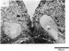
8. There were two types of synaptic ending on Müller cell bodies, one type containing round vesicles and the other containing ellipsoidal vesicles. These terminals were intermixed over the surface of the cell bodies and dendrites with no readily apparent segregation.
9. Intracellular records from the spinal axons of Müller cells during electrical stimulation of cranial nerves and spinal cord showed, in addition to the normal propagating action potential activity which normally originates in the cell bodies, depolarizing, hyperpolarizing and biphasic evoked potentials. These membrane responses were grossly similar in appearance to synaptic potentials except that the large depolarizing potentials had unusually long decay times. The physiological basis of these potentials remains unclear.
10. Electron microscopic examination showed very few synaptic endings afferent to Müller axons, a finding in contrast to the abundance of synaptic-like potentials recorded. However, the occasional synapses afferent to Müller axons were invariably located near an efferent synaptic region of the axon itself. This raises the possibility that a very limited number of synaptic regions of Müller axons may be subject to presynaptic modulation of transmitter release.
11. The observations reported here support the idea that Müller cells in lamprey are an important motor outflow from the brain and serve to coordinate the lamprey's trunk responses to external sensory stimulation.
Full text
PDF


Images in this article


Selected References
These references are in PubMed. This may not be the complete list of references from this article.
- BERTOLINI B. ULTRASTRUCTURE OF THE SPINAL CORD OF THE LAMPREY. J Ultrastruct Res. 1964 Aug;11:1–24. doi: 10.1016/s0022-5320(64)80089-7. [DOI] [PubMed] [Google Scholar]
- DUDEL J., KUFFLER S. W. Presynaptic inhibition at the crayfish neuromuscular junction. J Physiol. 1961 Mar;155:543–562. doi: 10.1113/jphysiol.1961.sp006646. [DOI] [PMC free article] [PubMed] [Google Scholar]
- Diamond J., Huxley A. F. The activation and distribution of GABA and L-glutamate receptors on goldfish Mauthner neurones: an analysis of dendritic remote inhibition. J Physiol. 1968 Feb;194(3):669–723. doi: 10.1113/jphysiol.1968.sp008432. [DOI] [PMC free article] [PubMed] [Google Scholar]
- FRANKENHAEUSER B., HODGKIN A. L. The action of calcium on the electrical properties of squid axons. J Physiol. 1957 Jul 11;137(2):218–244. doi: 10.1113/jphysiol.1957.sp005808. [DOI] [PMC free article] [PubMed] [Google Scholar]
- KUNO M. MECHANSIM OF FACILITATION AND DEPRESSION OF THE EXCITATORY SYNAPTIC POTENTIAL IN SPINAL MOTONEURONES. J Physiol. 1964 Dec;175:100–112. doi: 10.1113/jphysiol.1964.sp007505. [DOI] [PMC free article] [PubMed] [Google Scholar]
- Lowenstein O., Osborne M. P., Thornhill R. A. The anatomy and ultrastructure of the labyrinth of the lamprey (Lampetra fluviatilis L.). Proc R Soc Lond B Biol Sci. 1968 Jun 11;170(1019):113–134. doi: 10.1098/rspb.1968.0029. [DOI] [PubMed] [Google Scholar]
- Martin A. R., Wickelgren W. O., Ber1anek R. Effects of iontophoretically applied drugs on spinal interneurons of the lamprey. J Physiol. 1970 May;207(3):653–665. doi: 10.1113/jphysiol.1970.sp009086. [DOI] [PMC free article] [PubMed] [Google Scholar]
- Martin A. R., Wickelgren W. O. Sensory cells in the spinal cord of the sea lamprey. J Physiol. 1971 Jan;212(1):65–83. doi: 10.1113/jphysiol.1971.sp009310. [DOI] [PMC free article] [PubMed] [Google Scholar]
- RESTIEAUX N. J., SATCHELL G. H. A unitary study of the reticulomotor system of the dogfish, Squalus lebruni (Vaillant). J Comp Neurol. 1958 Jun;109(3):391–415. doi: 10.1002/cne.901090305. [DOI] [PubMed] [Google Scholar]
- Rovainen C. M., Johnson P. A., Roach E. A., Mankovsky J. A. Projections of individual axons in lamprey spinal cord determined by tracings through serial sections. J Comp Neurol. 1973 May 15;149(2):193–202. doi: 10.1002/cne.901490205. [DOI] [PubMed] [Google Scholar]
- Rovainen C. M. Physiological and anatomical studies on large neurons of central nervous system of the sea lamprey (Petromyzon marinus). I. Müller and Mauthner cells. J Neurophysiol. 1967 Sep;30(5):1000–1023. doi: 10.1152/jn.1967.30.5.1000. [DOI] [PubMed] [Google Scholar]
- Rovainen C. M. Synaptic interactions of reticulospinal neurons and nerve cells in the spinal cord of the sea lamprey. J Comp Neurol. 1974 Mar 15;154(2):207–223. doi: 10.1002/cne.901540207. [DOI] [PubMed] [Google Scholar]
- Smith D. S., Järlfors U., Beránek R. The organization of synaptic axcplasm in the lamprey (petromyzon marinus) central nervous system. J Cell Biol. 1970 Aug;46(2):199–219. doi: 10.1083/jcb.46.2.199. [DOI] [PMC free article] [PubMed] [Google Scholar]
- WOLSTENCROFT J. H. RETICULOSPINAL NEURONES. J Physiol. 1964 Oct;174:91–108. doi: 10.1113/jphysiol.1964.sp007475. [DOI] [PMC free article] [PubMed] [Google Scholar]