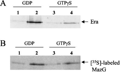
FIG. 2.
MazG and Era interaction in vitro. (A) Purified MBP-MazG fusion protein was mixed with Era in the presence of 20 μM GTPγS or GDP, and then the protein complexes were pulled down with amylose resin. The bound proteins on the resin were eluted, resolved on SDS-PAGE, blotted onto a polyvinylidene difluoride membrane, and detected with rabbit anti-Era antiserum. Lane 1, MBP, Era, and GDP; lane 2, MBP-MazG, Era, and GDP; lane 3, MBP, Era, and GTPγS; lane 4, MBP-MazG, Era, and GTPγS. (B) Purified MBP-Era fusion protein was mixed with in vitro-translated MazG labeled with [35S]Met in the presence of 20 μM GTPγS or GDP, and then the complexes were pulled down with amylose resin. The bound proteins on the resin were eluted and resolved on SDS-PAGE followed by autoradiography. Lane 1, MBP, [35S]MazG, and GDP; lane 2, MBP-Era, [35S]MazG, and GDP; lane 3, MBP, [35S]MazG, and GTPγS; lane 4, MBP-Era, [35S]MazG, and GTPγS.

