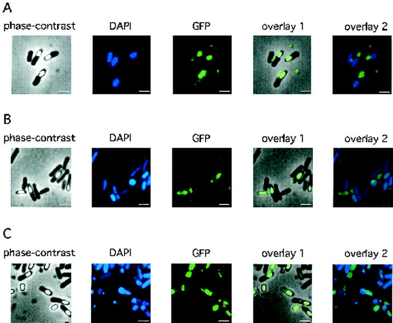
FIG. 5.
Localization of the PdaA-GFP fusion protein in the wild-type strain (A) or pdaA-deficient mutant YFJSd (B and C). The DAPI or GFP fluorescence of cells at t20 was detected as described in Materials and Methods. The pdaA-gfp-containing plasmid (pHYPRfjSG) was used for transformation of B. subtilis 168 or YFJSd. Spores and sporangia were collected at t20 at 37 and 30°C in DSM medium (B and C, respectively). The culture at 30°C exhibited stronger fluorescence in spores than that at 37°C. Overlay 1 is a phase-contrast image overlayed on a GFP image, and overlay 2 is a DAPI-stained fluorescence image overlayed on a GFP image. Bars, 2 μm.

