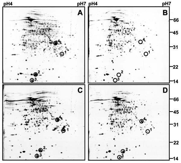
FIG. 3.
Two-dimensional gel electrophoretic analyses of protein extracts from wild-type BCG and ΔdosR::km, ΔdosR::km(pCB4), and ΔRv3132c::km strains grown in the Wayne dormancy culture system. Protein extracts were prepared at the time when cultures grown in the Wayne system terminated growth at day 5, as indicated by the arrow in Fig. 2A. Then 100-μg samples of total protein were subjected to two-dimensional gel electrophoresis. Silver-stained gels are shown. (A) Wild-type BCG. (B) BCG ΔdosR::km. (C) BCG ΔdosR::km(pCB4). Two protein spots that were detected in the pCB4-transformed ΔdosR::km strain upon termination of growth are marked by arrows; their identity remains to be determined. (D) BCG ΔRv3132c::km. Circles labeled 1 to 4 indicate dormancy-induced protein spots. In B, where the dormancy proteins were not detectable, the circles indicate their expected migration positions. 1, DosR; 2, α-crystallin; 3, conserved hypothetical protein Rv2626c; 4, conserved hypothetical protein Rv2623. Sizes are indicated in kilodaltons. The experiments were repeated twice with independently prepared cultures, yielding the same results. The experiments were also carried out for all four strains with protein extracts from 20-day-old Wayne cultures, yielding the same results that were obtained for 5-day-old cultures (data not shown).

