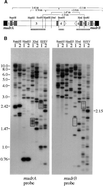
Figure 4.
Molecular Organization of Homologous MuDR Genes.
(A) Diagram of MuDR and the sizes of expected restriction fragments analyzed in (B). The hatched boxes represent DNA hybridization probes. Note that the mudrA probe is composed of a mixture of exon 3– and exon 4–specific fragments, as described in Methods.
(B) DNA samples from the bz2 non-Mutator, W23 inbred line (lanes 1), and the bz2-mu2 active Mutator line (lanes 2) were digested with restriction enzymes as described in (A). Sequential DNA gel blot analysis with mudrA- and mudrB-specific probes demonstrates hybridization of both probes to fragments of the same size, as appropriate, based on the restriction map of MuDR. Some of the most prominent cohybridizing bands (boxes) that differ in size from the wild-type MuDR span the repeat-rich intergenic region, for which length variation has already been reported (Gutiérrez-Nava et al., 1998). Other features are discussed in the text. EI/EV, EcoRI/EcoRV.

