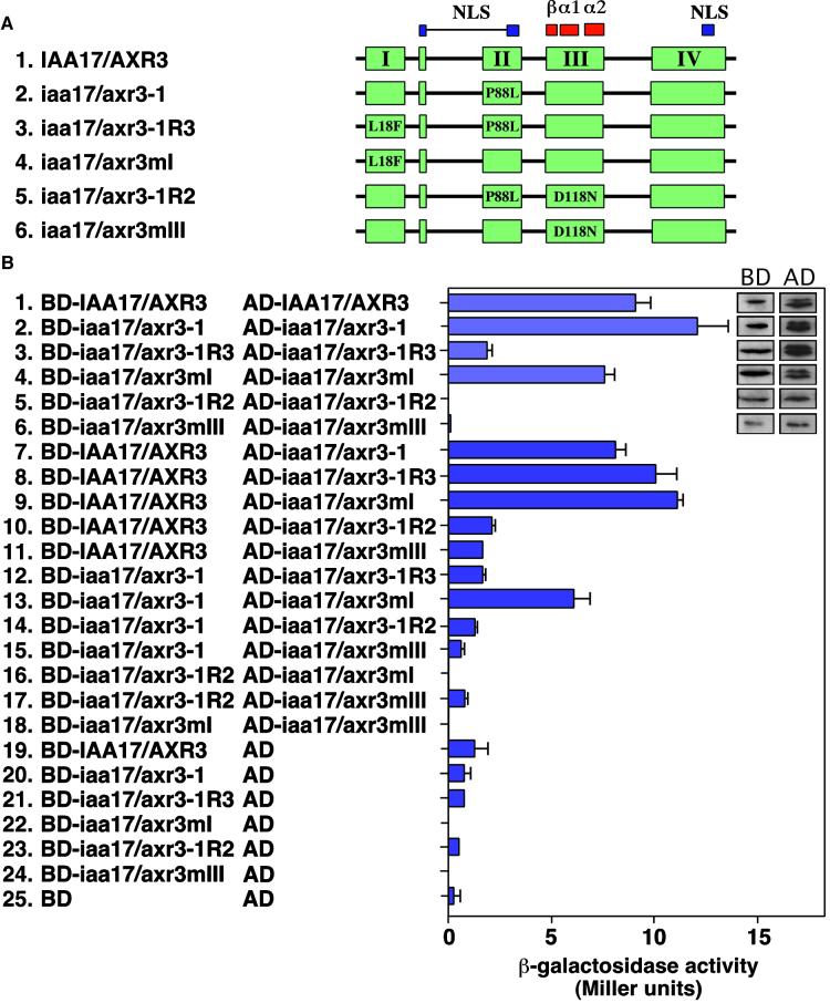
Figure 4.
Homodimerization of Wild-Type and Mutant Forms of IAA17/AXR3.
(A) Scheme of the constructs used. The coloring and designation of conserved elements are the same as in Figure 3A. The amino acid changes and their positions in the mutant proteins are indicated. The different cDNAs were fused in frame at the 3′ end of either the GAL4 AD or the GAL4 BD.
(B) Analysis of IAA17/AXR3 homodimerization using the yeast two-hybrid system. Y190 cells transformed with the indicated plasmids were analyzed for the level of β-galactosidase activity (Miller units). The two columns of inset sections show immunoblots demonstrating the expression of different forms of the IAA17/AXR3 protein expressed as fusions with either the GAL4 DNA BD or the GAL4 AD. The level of β-galactosidase activity was determined using orthonitrophenyl-β-d-galactopyranoside as a substrate. The values shown are averages of triplicate assays performed with at least two independent yeast colonies. Error bars represent the standard deviation. AD, plasmid with the GAL4 activation domain alone; BD, plasmid with the GAL4 binding domain alone.

