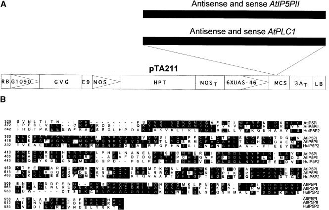
Figure 1.
Structures of the Transgenic Constructs and Alignment of the Ins(1,4,5)P3 5-Phosphatase Amino Acid Sequences.
(A) Structure of the pTA211 plasmid with the left and right T-DNA borders. AtPLC1 and AtIP5PII cDNAs were cloned in both the sense and antisense orientations into the multiple cloning site of the binary plasmid pTA211, which is a modification of the original pTA7001 vector (Aoyama and Chua, 1997) (see Methods). RB, right T-DNA border; G1090, G10-90 promoter (Ishige et al., 1999); GVG, glucocorticoid-inducible chimeric transcription factor; E9, pea rbcS-E9 polyadenylation sequence; NOS, nopaline synthetase promoter; HPT, hygromycin phosphotransferase coding sequence; NOST, nopaline synthetase polyadenylation sequence; 6XUAS-46, GVG-regulated promoter; MCS, multiple cloning site; 3AT, rbcS-3A polyadenylation sequence; LB, left T-DNA border.
(B) Alignment of the conserved amino acid sequences of the human Ins(1,4,5)P3 5-phosphatase type II (HuI5P2; Jefferson and Majerus, 1995) and Arabidopsis AtIP5PI and AtIP5PII. Identical amino acids are shaded in black. Alignment was performed using Clustal methods with the DNASTAR (Madison, WI) program.

