
Figure 6.
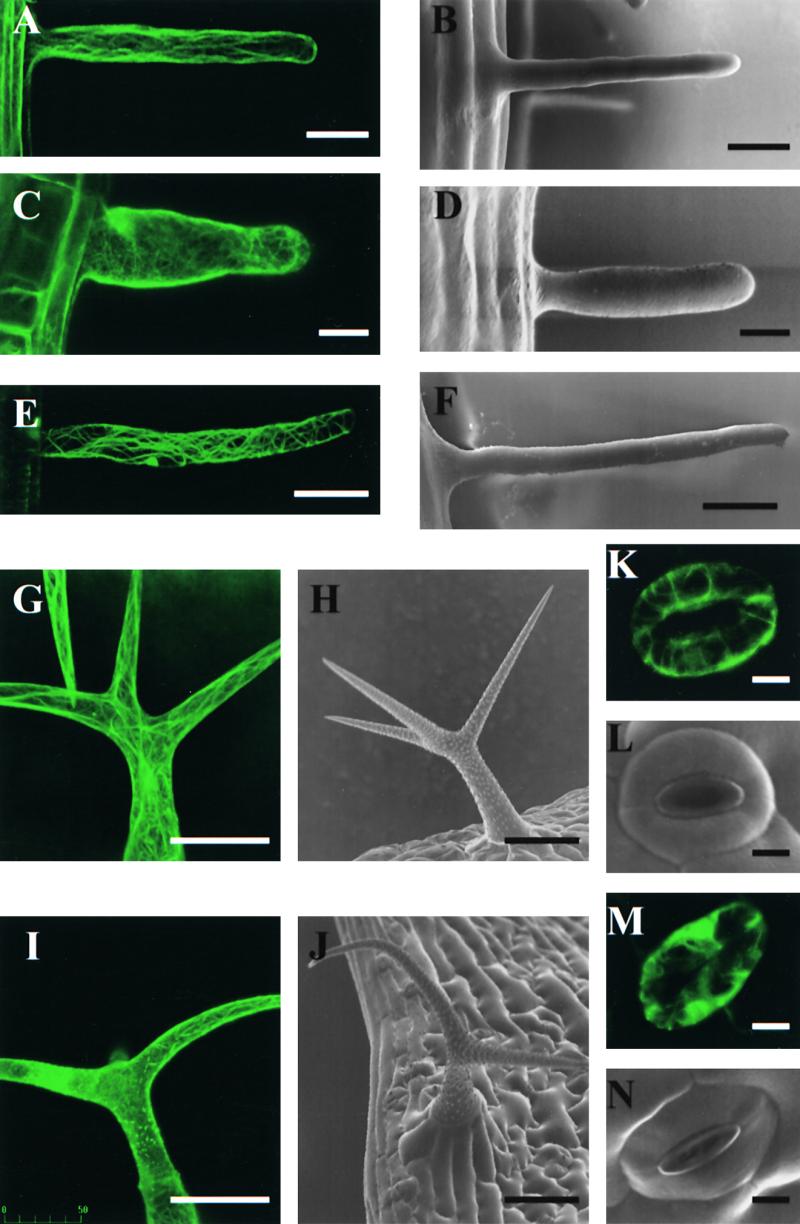
Morphology and Actin Cytoskeleton of Specialized Cell Types.
(B), (D), (F), (H), (J), (L), and (N) show cell morphology as observed using scanning electron microscopy; (A), (C), (E), (G), (I), (K), and (M) show F-actin organization as visualized using stable expression of GFP-mTn. Projections of serial confocal optical sections are shown. All images depict selected cells with representative phenotypes. Wild-type trichomes display a variable degree of branching (Mathur and Chua, 2000), which was not affected significantly by AtADF1 overexpression (data not shown).
(A) and (B) Wild-type root hair.
(C) and (D) AtADF1-O root hair.
(E) and (F) AtADF-U root hair.
(G) and (H) Wild-type trichome.
(I) and (J) AtADF1-O trichome.
(K) and (L) Wild-type stomata.
(M) and (N) AtADF1-O stomata.
 in (A) to (N)
  .
.