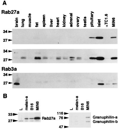
FIG. 2.
Tissue and cell expression of Rab27a, Rab3a, and granuphilin. (A) Isolation of mouse pancreatic islets with collagenase and preparation of mouse tissues and cell extracts were performed as described previously (37). An equal amount of protein (100 μg) was loaded onto a polyacrylamide gel. Immunoblotting was performed using anti-Rab27a (top panel) and anti-Rab3a (bottom panel) antibodies. A longer exposure of the Rab27a immunoblot is also shown (middle panel). (B) An equal amount of protein (30 μg) from mouse cell lines (L fibroblast, melan-a melanocyte, B16 melanoma, and MIN6 cells) was loaded onto a polyacrylamide gel. Immunoblotting was performed using anti-Rab27a (left panel) and antigranuphilin (αGrp-N, right panel) antibodies. Numbers to the left of each panel are molecular masses in kilodaltons.

