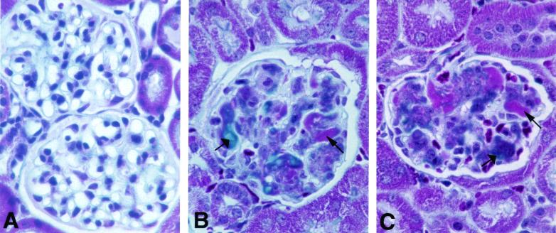
FIG. 4.
Glomerular lesions contain deposits of collagen and immune complexes. The composition of the lesions in the glomeruli of wild-type (A) and apoJ/clusterin-deficient (B and C) mice was examined by using the Masson-Trichrome stain. Blue staining (wide arrows) suggests the accumulation of collagen deposits in the glomeruli, whereas fuchsin staining (narrow arrows) indicates the deposition of immune complexes. Magnification, ×360.

