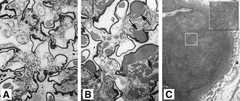
FIG. 5.
Electron-dense tubulo-fibrillar aggregates in the mesangium of apoJ/clusterin-deficient glomeruli. Sections from wild-type (A) and mutant (B) mice were silver stained. Mutant mice exhibited mesangial expansion and deposits of electron-dense material. Magnification, × 2,828. (C) High-power view of the mesangial deposits from mutant mice, demonstrating the tubulo-fibrillary structures. Magnification, ×18,800. The box indicates a region where the tubulo-fibrillary structures are evident.

