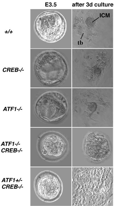
FIG. 3.
Functional disruption of both CREB and ATF1 results in preimplantation defects. Embryos of different genotypes were isolated at E3.5. ATF1+/− CREB−/− and ATF1−/− CREB−/− embryos show a morula-like structure, while wild-type and single-knockout embryos are comprised of trophectoderm surrounding ICM and blastocoel. Outgrowths after 4 days of in vitro culture of wild-type, ATF1−/−, and CREB−/− blastocysts develop a monolayer of trophoblast cells with the ICM cells on top. No outgrowth of ATF1−/− CREB−/− embryos occurs after 4 days in culture. Embryos seem to arrest in development; cells appear necrotic and are still surrounded by the zona pellucida. ATF1+/− CREB−/− embryos hatch from the zona pellucida; a trophoblast monolayer is present, but no obvious ICM can be seen. tb, trophoblast monolayer. 3d, 3 days.

