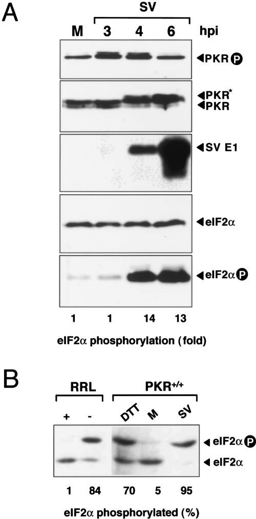
Figure 1.
PKR activation and eIF2α phosphorylation in SV-infected cells. (A) 3T3 cells were infected with SV at an MOI of 25 PFU/cell. At the indicated times, cell extracts were made and analyzed by Western blot using the indicated antibodies. Bands corresponding to viral structural proteins E1, PKR, phospho-PKR, eIF2α, and phopho-eIF2α are shown. (Lower panel) The ratio of phosphorylated versus total eIF2α was estimated by densitometry of corresponding bands. (B) IEF analysis of eIF2α phosphorylation. 3T3 cells were SV-infected (4 h) or treated with 1 mM DTT (1 h), then analyzed by IEF (see Materials and Methods). Mock-infected cells (M) were included, as well as RRL-treated (+) or untreated (–), with hemine and EDTA as negative and positive controls of eIF2α phosphorylation, respectively. Phosphorylated and unphosphorylated eIF2α forms were quantified by densitometry.

