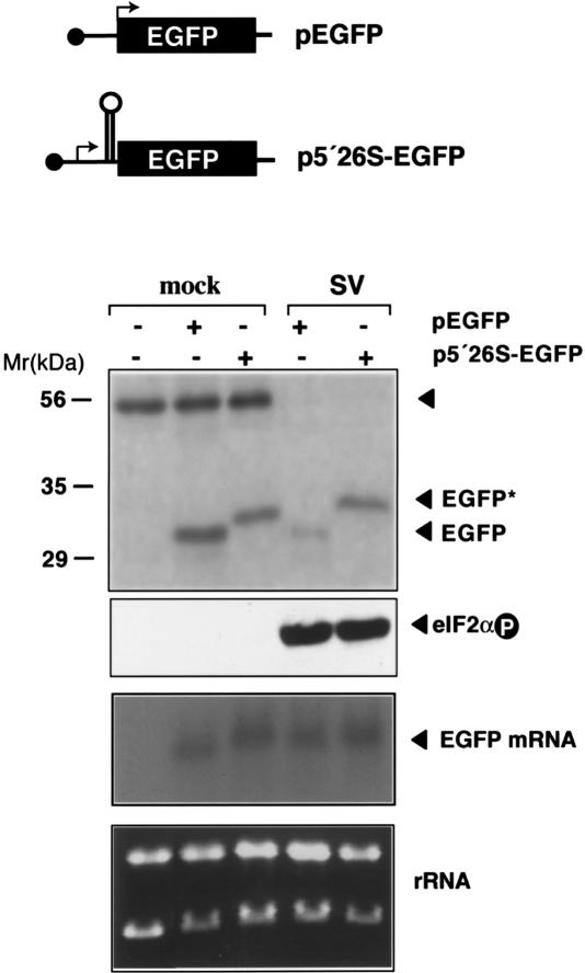
Figure 6.
Translation resistance to eIF2α phosphorylation promoted by the 5′ extreme of SV 26S mRNA. Diagram of EGFP constructs. p5′26S-EGFP contains the first 140 nt of SV before the EGFP-coding sequence. Arrows indicate translation initiation sites. BHK-21 cells were transfected with 2 μg of the indicated plasmids using JetPEI (Poly-Plus Transfection) and infected (SV) or not (mock) 48 h later with SV (MOI: 25 PFU/cell). (Upper panel) At 5 hpi, cells were labeled with [35S]Met/Cys (1 h) and immunoprecipitated with anti-EFGP antibodies. The autoradiogram of labeled products is shown. The protein band that cross-precipitated with anti-EGFP antibodies probably corresponds to actin and serves as an internal control. Western blot analysis of eIF2α phosphorylation and Northern blot analysis of EGFP mRNA levels are also shown (middle panel), as well as ethidium bromide staining of total RNA loaded in each sample (bottom panel). For Northern blot analysis, the membrane was probed with a 32P-labeled DNA fragment corresponding to the first 600 nt of the EGFP gene.

