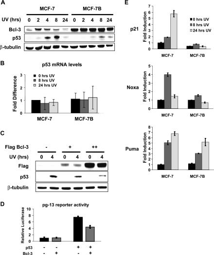
Figure 2.
Bcl-3 overexpression inhibits DNA damage-induced p53 activity. (A) UV-induced p53 protein levels are reduced in MCF-7B cells. MCF-7 and MCF-7B cells were treated with 40 J/m2 UV-C for the indicated times, and Western analysis was performed on whole-cell extracts using antibodies against Bcl-3, p53, and β-tubulin. (B) Overexpression of Bcl-3 does not affect p53 mRNA levels. MCF-7 and MCF-7B cells were treated with 40 J/m2 UV-C for the indicated times, and relative expression of p53 was measured by quantitative real-time PCR. (Lane 1) Expression levels were normalized to expression of glucuronidase-β, and the values represent the fold increase or decrease relative to untreated MCF-7 cells. (C) Transient expression of Bcl-3 leads to decreased p53 protein levels following UV treatment. MCF-7 cells were transfected with either empty vector or 2 or 4 μg of pCMV-Flag-Bcl-3. Two days after transfection, the cells were left untreated or were treated with 50 J/m2 UV-C for 4 h, and Western analysis was performed with antibodies against the Flag epitope, p53, or β-tubulin. (D) Transient expression of Bcl-3 inhibits p53 transcriptional activity. MCF-7 cells were transfected with 50 ng of pg-13-luciferase and 5 ng of renilla luciferase plus 100 ng of pCMV-Flag-Bcl-3 and pCMV-Flag-p53 where indicated. Firefly luciferase activity was measured and normalized to renilla luciferase. (Lane 1) Values represent fold increase over basal activity. (E) DNA damage-induced expression of p53 target genes is lost in MCF-7B cells. MCF-7 and MCF-7B cells were treated with 40 J/m2 UV-C for the indicated times, and relative expression of p21, Noxa, and Puma was measured by quantitative real-time PCR. (Lane 1) Expression levels were normalized to expression of glucuronidase-β, and the values represent the fold increase or decrease relative to untreated MCF-7 cells.

