Figure 7.
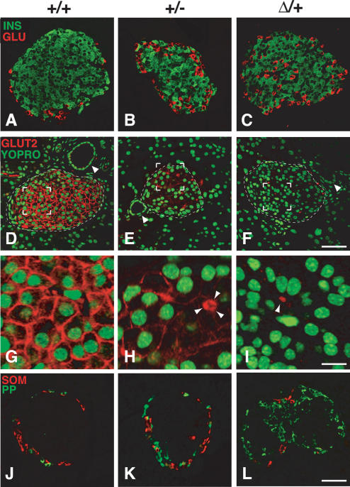
The Pdx1ΔI-II-III/+ genotype leads to severely compromised mature islet architecture and endocrine differentiation. All tissues analyzed were from 4-wk-old mice. (A-C) Pdx1ΔI-II-III/+ (Δ/+) mice show a reproducible increase and intra-islet scattering of glucagon cells (see also Supplementary Fig. 3) (D-F) β-Cell-selective expression of Glut2 is modestly decreased in Pdx1+/- (+/-) animals, but is absent in Δ/+ animals (arrowheads indicate ducts). (G-I) High magnification of bracketed region in D-F (arrowheads in H,I indicate autofluorescent erythrocytes in capillaries to show gain setting equivalence across the three panels; three erythrocytes clustered in H). (J-L) Frequency of PP cells is dramatically increased in Δ/+ animals; somatostatin cells are relatively unchanged in number and location (see also Supplementary Fig. 3). Bars: A-F, 50 μm; G-I, 12.5 μm; J-L, 50 μm.

