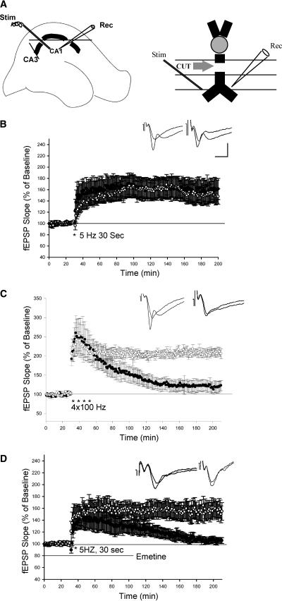
Figure 2.
LTP induced by theta frequency stimulation is unimpaired in isolated dendrites. (A) Schematic diagram showing the two incisions made to isolate CA1 dendrites and showing the placement of recording (Rec) and stimulating electrodes (Stim). (B) LTP induced by theta frequency stimulation in isolated dendrites is unimpaired. (○) LTP in the intact slices; (•) LTP in the isolated dendrites. (C) LTP induced by four trains of 100-Hz stimulation is reduced in isolated dendrites. (○) L-LTP in intact slices; (•) L-LTP in isolated dendrites. (D) Application of emetine (100 μM) blocked late phase of theta LTP in isolated dendrites. (○) Control; (•) emertine-treated slices. Sample traces before and 3 h after LTP are shown in the insets. Calibration: 2 mV, 10 ms.

