
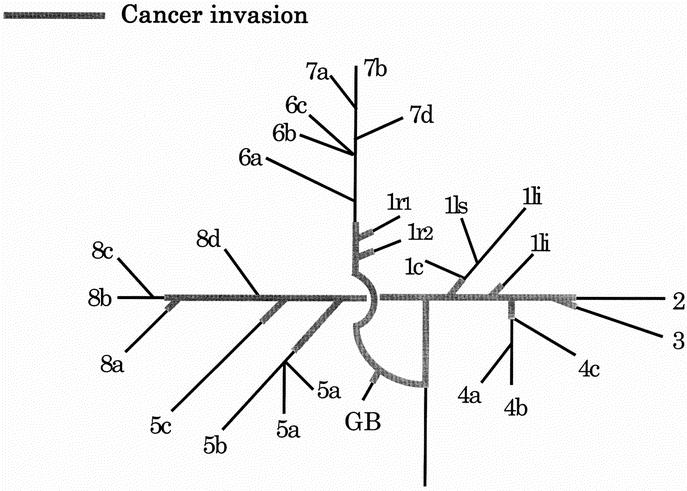
FIGURE 1. Two-dimensional map of the confluence patterns of intrahepatic and extrahepatic bile ducts and cancer extension along the bile ducts (the “pressed flower method”). Numbers refer to the Couinaud’s segment. 1li, left inferior branch; 1ls, left superior branch; 1c, caudate process branch; 1r, right branch; 4a, inferior branch; 4b, superior branch; 4c, dorsal branch; 5a, ventral branch; 5b, dorsal branch; 5c, lateral branch; 6a, ventral branch; 6b, dorsal branch; 6c, lateral branch; 7a, ventral branch; 7b, dorsal branch; 7d, medial branch; 8a, ventral branch; 8b, lateral branch; 8c, dorsal branch; 8d, medial branch; and GB, gallbladder.