
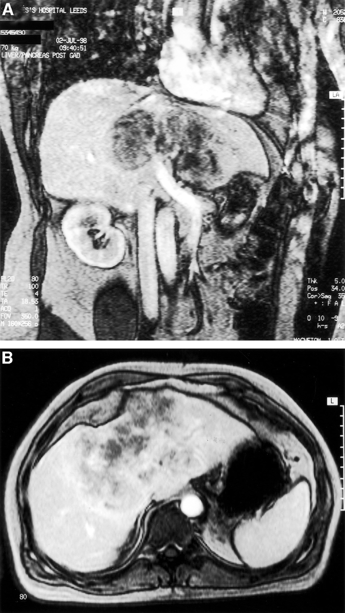
FIGURE 3. Coronal (A) and transverse (B) section magnetic resonance imaging demonstrates a 12 × 10-cm tumor enveloping both left and right branches of the portal vein, hepatic artery, and hepatic ducts (patient 4), for which an orthotopic liver transplant was performed.