
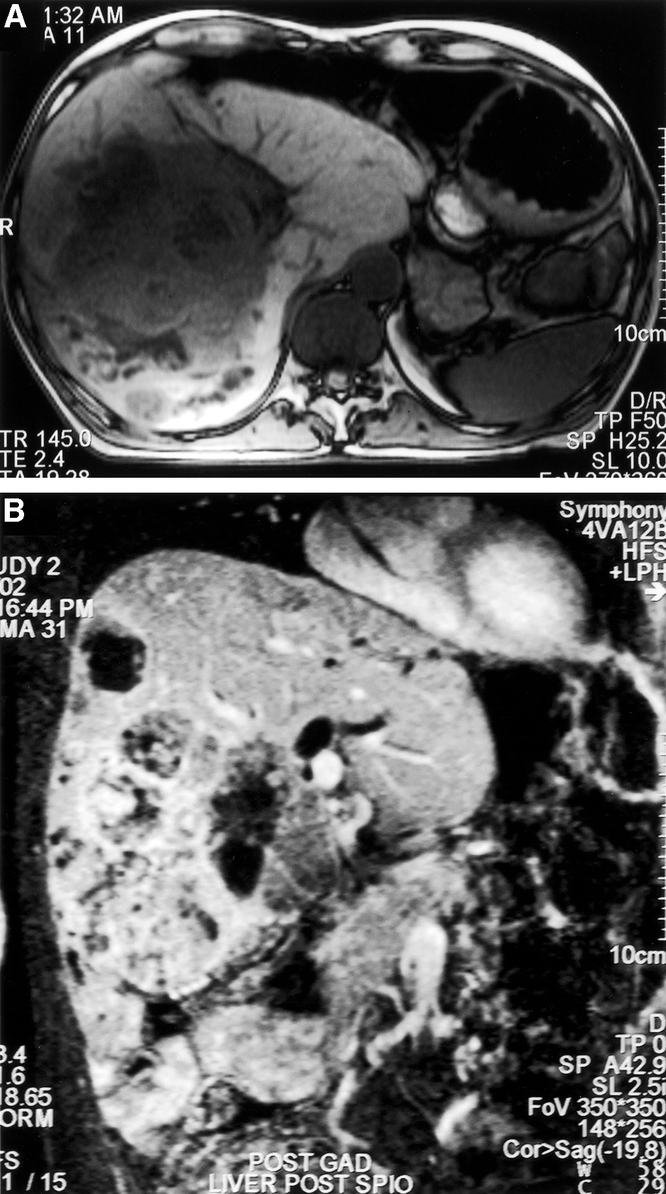
FIGURE 4. Transverse (A) and coronal oblique (B) section magnetic resonance imaging demonstrating a 15-cm diameter tumor, primarily involving the right liver, but with significant extension into the left liver. There is evident biliary obstruction (patient 7). Hepatic resection was possible by a modified right hemihepatectomy technique.