
Figure 4.
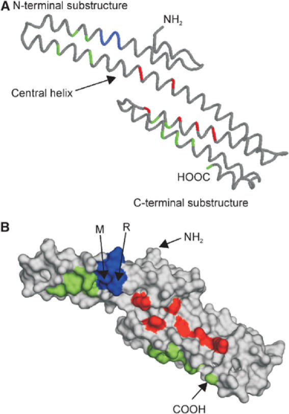
Monomeric E2 domain/central APP domain (CAPPD) (PDB:1RW6). (A) Backbone diagram: position of RERMS sequence is marked in blue, amino-acid residues forming the two hydrophobic patches are colored in green and the conserved amino-acid residues of the HSPG-binding site in red. (B) Surface representation of (A). The N- and C-termini as well as the underlined amino-acid residues of the RERMS sequence, which participate in dimerization, are labeled.