
Figure 1.
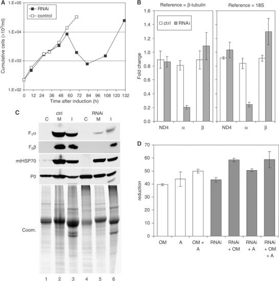
Knockdown of ATP synthase subunit α expression is lethal to LS stage T. brucei. (A) Culture growth shown as cumulative cell numbers after normalization for dilution during cultivation. Expression of subunit α was silenced using tet-inducible RNAi (solid squares); uninduced cells were maintained in parallel (open squares). (B) Determination of mRNA levels for ATP synthase subunits α and β and for the mitochondrial transcript ND4 by real-time RT–PCR (ΔΔCt method). Total RNA was isolated from induced cells after 43 h as well as from uninduced control cells. Primers for subunit α were located outside the region targeted by RNAi. Relative changes in mRNA levels with respect to the parental cell line are indicated, using β-tubulin mRNA or 18S rRNA for normalization. Average numbers for four amplifications are shown, using RNA preparations from two independent RNAi experiments. (C) Western blot analysis of crude cytosolic (C), soluble mitochondrial (M), and insoluble (I) fractions prepared from RNAi-induced cells after 45 h and from uninduced control cells. The blots were probed with reagents detecting the indicated proteins. Coomassie staining (bottom panel) revealed the protein loading. (D) Analysis of ATPase activity in crude mitochondrial fractions, generated as in (C) and assayed by measuring release of free phosphate. ATP synthase inhibitors oligomycin (OM; 2.5 μg/ml) and azide (A; 1 mM) were added where indicated. Average numbers for four assays are shown, using extract preparations from two independent RNAi experiments.