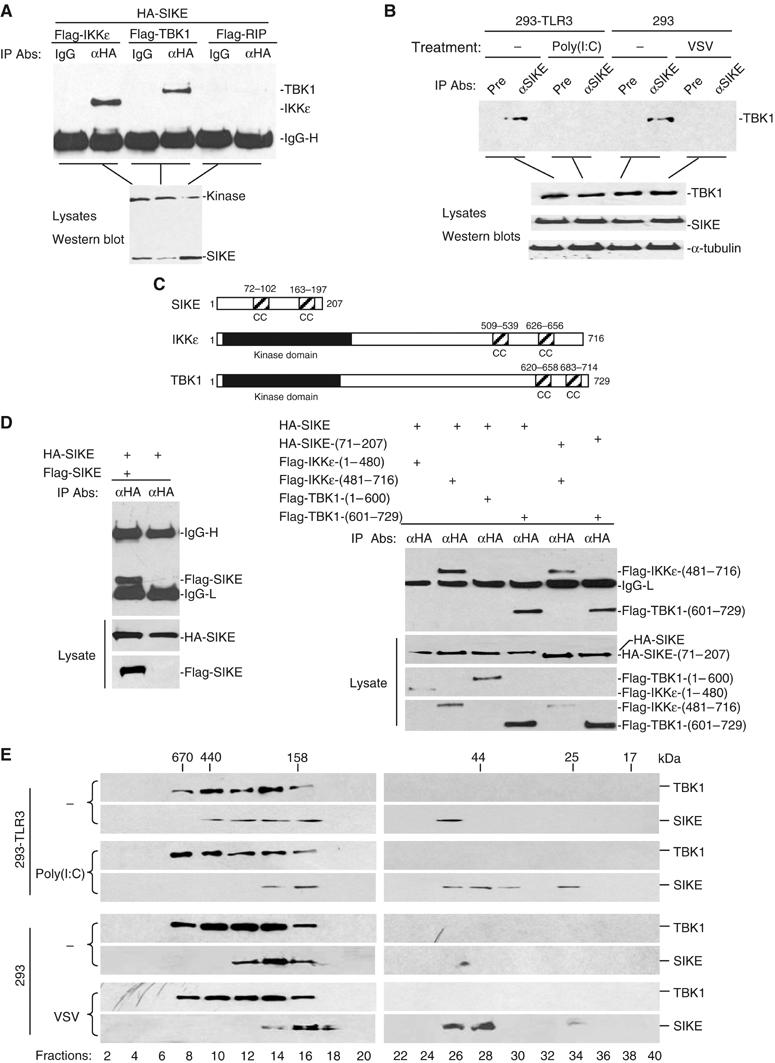
Figure 2.

SIKE is associated with IKKɛ and TBK1. (A) SIKE interacts with IKKɛ and TBK1 but not RIP in mammalian overexpression system. The 293 cells (1 × 10⁶) were transfected with the indicated plasmids (5 μg each). Co-immunoprecipitations were performed with anti-HA antibody (αHA) or control mouse IgG (IgG). Western blot was performed with anti-Flag antibody (upper panel). Expression of the transfected plasmids was confirmed by Western blot analysis of the lysates with anti-HA and anti-Flag antibodies (lower panel). (B) Association of SIKE with TBK1 and effects of stimulation on the association in untransfected cells. The 293-TLR3 or 293 cells (5 × 10⁷) were left untreated or treated with poly(I:C) (50 μg/ml) for 10 min or infected with VSV for 4 h. The cells were lysed and the lysates were immunoprecipitated with anti-SIKE antiserum or preimmune control serum. The immunoprecipitates were analyzed by Western blots with anti-TBK1 antibody (upper panel). The expression levels of the endogenous proteins were detected by Western blot analysis with anti-TBK1, anti-SIKE and anti-α-tubulin antibodies (lower panels). (C) A schematic presentation of the coiled-coil motifs identified in SIKE, IKKɛ and TBK1. CC, coiled coil. (D) SIKE interacts with itself, IKKɛ and TBK1 through their respective coiled-coil motifs. The 293 cells (1 × 10⁶) were transfected with the indicated plasmids (5 μg each) and co-immunoprecipitations were performed as in (A). (E) Analysis of protein complexes containing SIKE and TBK1 by size-exclusion chromatography. The 293-TLR3 (2 × 10⁸) cells were treated with poly(I:C) (50 μg/ml) for 10 min or left untreated. The 293 cells were infected with VSV for 4 h or left uninfected before lysis. Cell lysates were analyzed by size-exclusion chromatography on Superdex 200 column. The individual fractions were analyzed by Western blots with anti-TBK1 and anti-SIKE antibodies, respectively.
