
Figure 2.
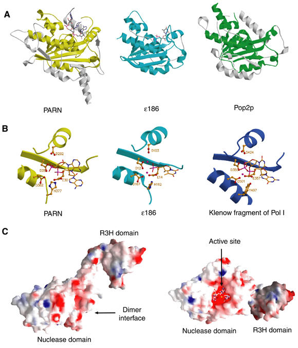
Comparison of PARNn with other members of the DEDD family. (A) Structural comparison of the nuclease domain of PARNn with those of ɛ186 and Pop2p. The DEDD core domains are colored yellow, cyan and green for PARN, ɛ186 and Pop2p, respectively, with the rest of the molecules colored in pale gray. Bound nucleotides are shown in stick model. (B) Structures of the active sites of PARNn, ɛ186 of Pol III and the klenow fragment of Pol I. Bound nucleotides are shown in stick model, catalytic residues in ball-and-stick model and metal ions in CPK model colored with magenta. (C) Solvent-accessible and electrostatic potential of PARNn colored from blue (basic) to red (acidic). For simplicity, only one subunit is shown. Left panel: the side view of the electrostatic potential surface. Right panel: the top view of the surface rotated about 90° around y axis relative to the view in the left panel.