
Figure 6.
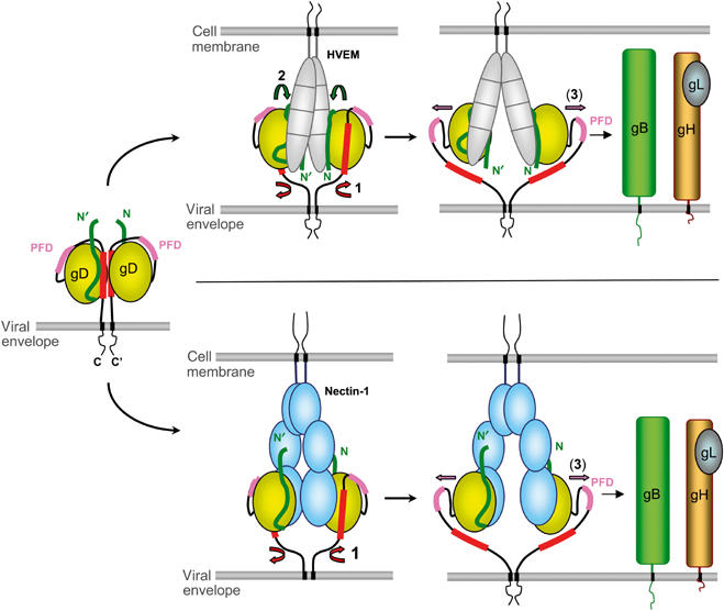
Proposed mechanism for receptor-mediated activation of HSV gD. Envelope gD is shown, as a putative dimer, in its unbound state as well as during interaction with HVEM (top) or nectin-1 (bottom). Conformational changes are chronologically indicated by numbered arrows: (1) displacement of the C-terminus, (2) folding of the gD N-terminus in the case of HVEM binding, and (3) exposure of the PFD. The N-terminus of gD is shown in green, the C-terminus (290–299) is colored red, and the PFD (260–285) is pink.