
Figure 4.
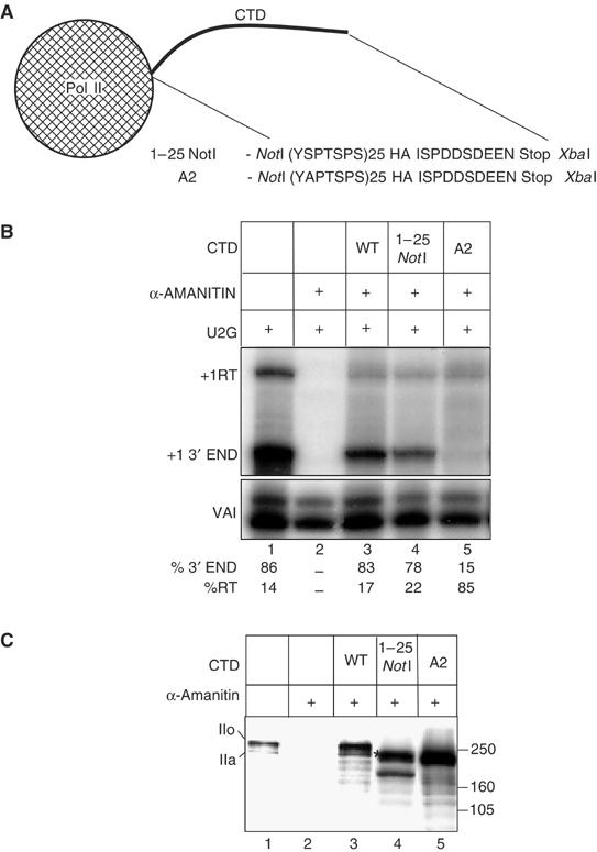
Mutation of Ser2 of the pol II CTD repeats causes readthrough of the U2 3′ box. (A) Diagram of the CTD region of the 1–25 NotI and A2 cassette constructs of the pol II large subunit showing the amino acids encoded by the region between the NotI and XbaI sites. HA and Stop denote the HA epitope tag and a stop codon respectively. (B) RNase protection analysis of RNA transcribed from U2G after ectopic expression of the α-amanitin resistant pol II large subunit constructs indicated above the lanes and treatment of cells with α-amanitin. The positions of the protected products are shown on the left. The relative amount of correct 3′ end and readthrough is noted below each lane. (C) Western blot analysis of the proteins encoded by the pol II large subunit constructs using an antibody against the large subunit of pol II (Santa Cruz, H-224). The construct transfected is shown above each lane. The position of the hyperphosphorylated form (IIo) and the hypophosphorylated form (IIa) of the endogenous and ectopically expressed pol II large subunits with a full-length CTD are noted on the left. The putative hyperphosphorylated form of 1–25 NotI is marked with an *.