
Figure 6.
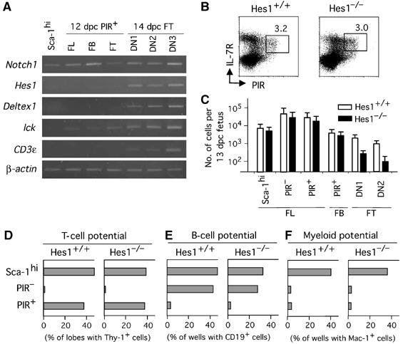
PIR+ T-cell progenitors in FL and FB emerge independently of Hes1. (A) RT–PCR analysis for expression of Notch-signal-related genes in cells of various stages of T-cell development. (B) PIR+ population in FL from 13 dpc fetuses of Hes1−/− mice. Cells were four-color stained in the same way as in Figure 1A, and profiles of Lin−c-kit+ cells are shown. (C) Numbers of Sca-1hi, PIR− and PIR+ cells of FL, PIR+ cells of FB, and DN1 and DN2 cells of FT from 13 dpc fetuses of Hes1−/− mice and Hes1+/+ littermate mice, are shown. Error bars indicate SD in each group. (D–F) PIR+ cells in FL from Hes1−/− fetuses are restricted to the T-cell lineage. Sca-1hi cells, PIR− cells and PIR+ cells of 13 dpc FL (a total of 100 cells for each group) from wild-type mice and Hes1−/− mice were examined for T, B and myeloid cell potential by coculture with a dGuo lobe for detection of T-cell potential (D), coculture with TSt-4 for B-cell potential (E), and coculture with PA6 for myeloid cell potential (F), respectively, except that Thy-1 was used as a marker for the T-lineage cells in (D).