
Figure 2.
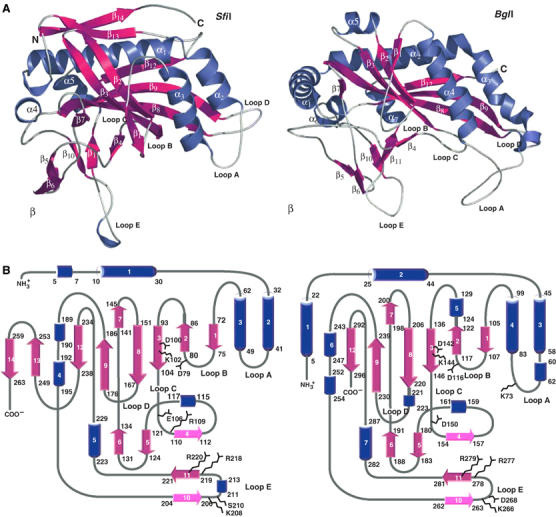
Comparisons between SfiI and BglI subunits. (A) Structure of SfiI (left) and BglI (right) monomers. The secondary structural elements are labeled and colored as follows: α helices are blue, β strands are magenta, and loops are white. The 3/10 helices are unlabeled. (B) Topology diagram of SfiI (left) and BglI (right) subunit. The color scheme is the same as in (A).