
Figure 6.
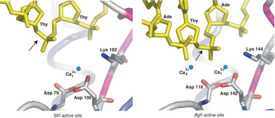
The active sites of SfiI (left) and BglI (right). The DNA and the active site residues of SfiI (Asp 79, Asp100, and Lys102) and BglI (Asp116, Asp142, and Lys144) are shown in ‘stick' representation. The scissile phosphodiester is indicated by arrows. The Ca2+ ions are shown as nonbonded spheres and colored aqua, water molecules are not shown. In SfiI, the second Ca2+ is missing and the scissile phosphodiester is ∼3 Å further away than in BglI, indicating that the enzyme is in an inactive state.