
Figure 3.
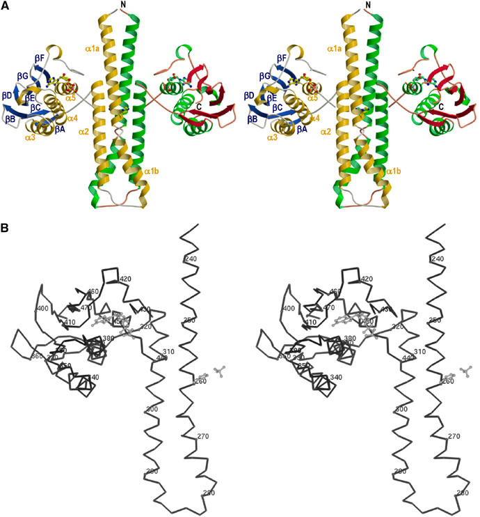
Molecular structure of the cytoplasmic portion of TM0853. (A) Ribbon representation of the crystallographic dimer of HK853–CD, including ADPβN. The α helices are labeled α1–α5, and colored gold (subunit A) and green (subunit B), β strands are labeled βA–βF, and colored blue (subunit A) and red (subunit B). The positions of N and C termini are labeled in subunit B. The ADPβN molecule and the phospho-acceptor residue (His260) are shown in ball-and-stick representation. The membrane would be located on the top of N-terminal residues. (B) Stereo Cα trace of the gold and blue protomer. Every tenth Cα is indicated as a sphere and numbered. The ADPβN molecule, the His260 and the sulfate ion coordinated with His260 are drawn in a gray ball-and-stick representation. The orientation is as in panel (A).