
Figure 4.
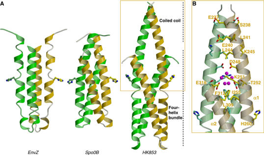
Comparison of DHp domains of EnvZ HK, Spo0B phosphotransferase and TM0853 HK. (A) Ribbon diagrams of the three DHp domains in their dimeric forms (protomers colored in green and gold). The DHp domains have been oriented with the plane containing the phospho-accepting histidines, which are shown in stick representation, and the principal helix axes parallel to the page. (B) Detail of the HK853 coiled-coil motif. Residues interacting in this motif are shown as stick representation and labeled on one protomer. Solvent molecules (magenta) are shown in the cavity generated at the juncture between the coiled coil of α1a helices and the four-helix bundle.