
Figure 5.
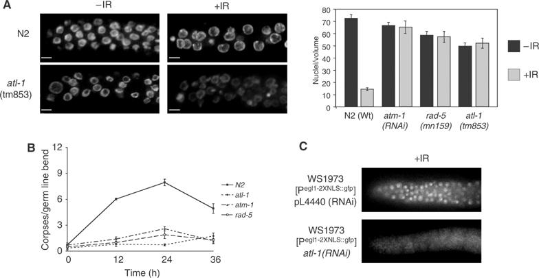
atl-1 is required for checkpoint-dependent cell cycle arrest and apoptosis following IR via the cep-1/egl-1 proapoptotic pathway. (A) Representative images of a single focal plane through the mitotic region of the germline from animals of the indicated genotype counterstained with DAPI following IR-treatment (scale bar=5 μm). The graph on the right shows quantification of IR-induced cell cycle arrest of mitotic germline nuclei in animals of the indicated genotype that was determined by scoring the number of nuclei in a volume of 54 000 μm3 12 h after exposure of L4 stage animals to 75 Gy IR, as previously described (Gartner et al, 2000). Error bars indicate standard error of mean from at least ten germlines for each experiment. (B) Germ cell apoptosis was measured by differential interference contrast (DIC) microscopy in animals of the indicated genotype at the indicated time points post IR-treatment. Error bars indicate standard error of mean from at least ten germlines for each time point. (C) Representative images of GFP expression from the Pegl-1::gfp transcriptional reporter (Hofmann et al, 2002) 30 h after IR treatment (120 Gy) in animals subjected to L4440 RNAi (control) and atl-1(RNAi).