Figure 1.

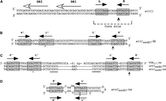
Integron recombination sites. (A) Sequence of the ds attI1 site. (B) Sequence of the ds attCaadA7 site. (C) Multiple sequence alignment of the attC sites bs studied in this work. (D) Proposed secondary structure for the attCaadA7 bs. The inverted repeats L, L′ and L″, R, R′ and R″ are indicated with black arrow; the asterisk (*) shows the position of the protruding G present in L″ relative to L′. The attI1 direct repeats bound by InI1 are indicated by horizontal lines with an empty arrowhead (Collis et al, 1998; Gravel et al, 1998a). The putative IntI1 binding domains, as defined by Stokes et al (1997), are marked with gray boxes. Vertical arrows indicate crossover position. The secondary structure was determined using the MFOLD (Walter et al, 1994) online interface at the Pasteur Institute.
