Figure 7.

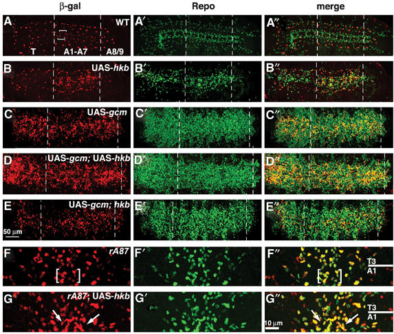
Role of Hkb and Gcm in NGB1-1A specification. (A–E″) Ventral views of stage 16 embryonic ventral cord carrying the P101 SPG marker; T3 and A1 indicate, respectively, third thoracic and first abdominal segments; anterior to the top and vertical line indicates the midline. β-gal (SPG), Repo double labeling. Left panels show β-gal, mid panels, Repo and right panels, merges. In all overexpression experiments, sca-GAL4 was used as a driver. (A–A″) labeling in wild-type (WT) embryo. Square brackets indicate A- and B-SPG (note that LV-SPG cannot be seen in this focal plane), arrowhead in (A) indicates lateral SPG, a cell that flanks A- and B-SPG cells, but is not derived from NGB1-1A lineage. (B–B″) labeling upon hkb overexpression induces additional SPG (B) and Repo (B′) labeling, close to the position at which A- and B-SPG are normally present. (C–C″) labeling upon combined gcm and hkb overexpression. Note the presence of additional SPG labeling in thoracic and abdominal segments (thick arrow). (D–D″) labeling upon gcm expression. (E–E″) labeling upon gcm expression in hkb embryo. (F–G″) Ventral views of stage 16 ventral cord of rA87/+ embryos. β-gal (rA87), Repo double labeling as above. (F–F″) labeling in a WT rA87/+ embryo. (G–G″) labeling in a sca-GAL4/rA87; UAS-hkb/+ embryo. Square brackets in (F and F″) indicate β-gal/Repo positive cells at the position of A- and B-SPG. Colocalization of additional Repo and β-gal labeling is indicated by arrows. Scale bar: 10 μm.
