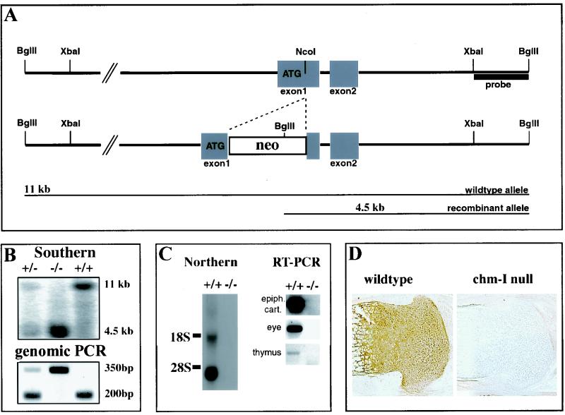
FIG. 1.
Targeted disruption of mouse chm-I. (A) Structure of wild-type allele, targeting construct, and recombinant locus. Boxes, exons. The expected fragment sizes after BglII digestion are 11 kb for the wild-type allele and 4.5 kb for the recombinant allele. (B) (Top) Southern blot analysis of mouse tail DNA isolated from the progeny of a mating between heterozygous parents. DNAs were digested with BglII and hybridized with the probe indicated in panel A. (Bottom) PCR genotyping with primers for wild-type and recombinant alleles. +/+, wild-type mouse; +/−, heterozygous mouse; −/−, homozygous mutant mouse. (C) (Left) Northern blot analysis of total RNA from limb cartilage derived from newborn wild-type and homozygous mutant mice. The filter was hybridized with a cDNA probe specific for chm-I. No RNA is detectable in the mutant mice. (Right) RT-PCR with chm-I-specific primers, cartilage, thymuses, and whole eyes of homozygous mutant and homozygous wild-type mice gives no evidence for chm-I mRNA in the chm-I-deficient mouse. (D) Immunohistochemistry of newborn tibial growth plate with a chm-I-specific antibody confirms complete deletion of the chm-I protein in the mutant mouse.

