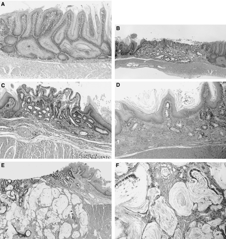
FIGURE 3. Photomicrographs showing esophageal histopathology. (Hematoxylin and eosin) A, Basal cell hyperplasia in the midesophagus of a rat from the DER20 group (×100). B, BE in a rat from the DER30 group. Columnar epithelium consists of absorptive cells with brush borders and goblet cells (×40). C, BE in a rat from the BD30 group (×100). D, Columnar cells under generating squamous epithelium are seen in a rat from the BD20 group (×100). E and F, Mucinous esophageal adenocarcinoma characterized by malignant glands associated with lakes of extracellular mucin in a rat from the DER50 group. The tumor is invading the adventitia (E, ×40; F, ×100). DER20, total gastrectomy and esophagojejunostomy to induce duodenoesophageal reflux, killed after 20 weeks; DER30, total gastrectomy and esophagojejunostomy to induce duodenoesophageal reflux, killed after 30 weeks; BD30, conversion from esophagojejunostomy to biliary diversion procedure to prevent duodenoesophageal reflux at the 30th week, killed after 50 weeks; BD20, conversion from esophagojejunostomy to biliary diversion procedure to prevent duodenoesophageal reflux at the 20th week, killed after 50 weeks; DER50, total gastrectomy and esophagojejunostomy to induce duodenoesophageal reflux, killed after 50 weeks.
